SATDI-IC – Field Emission Scanning Electron Microscope, FESEM (Sigma 300 VP)
Equipamiento
|
Microscopio electrónico de barrido de emisión de campo Sigma 300 VP (Zeiss) |
Campus de ELCHE |
 |
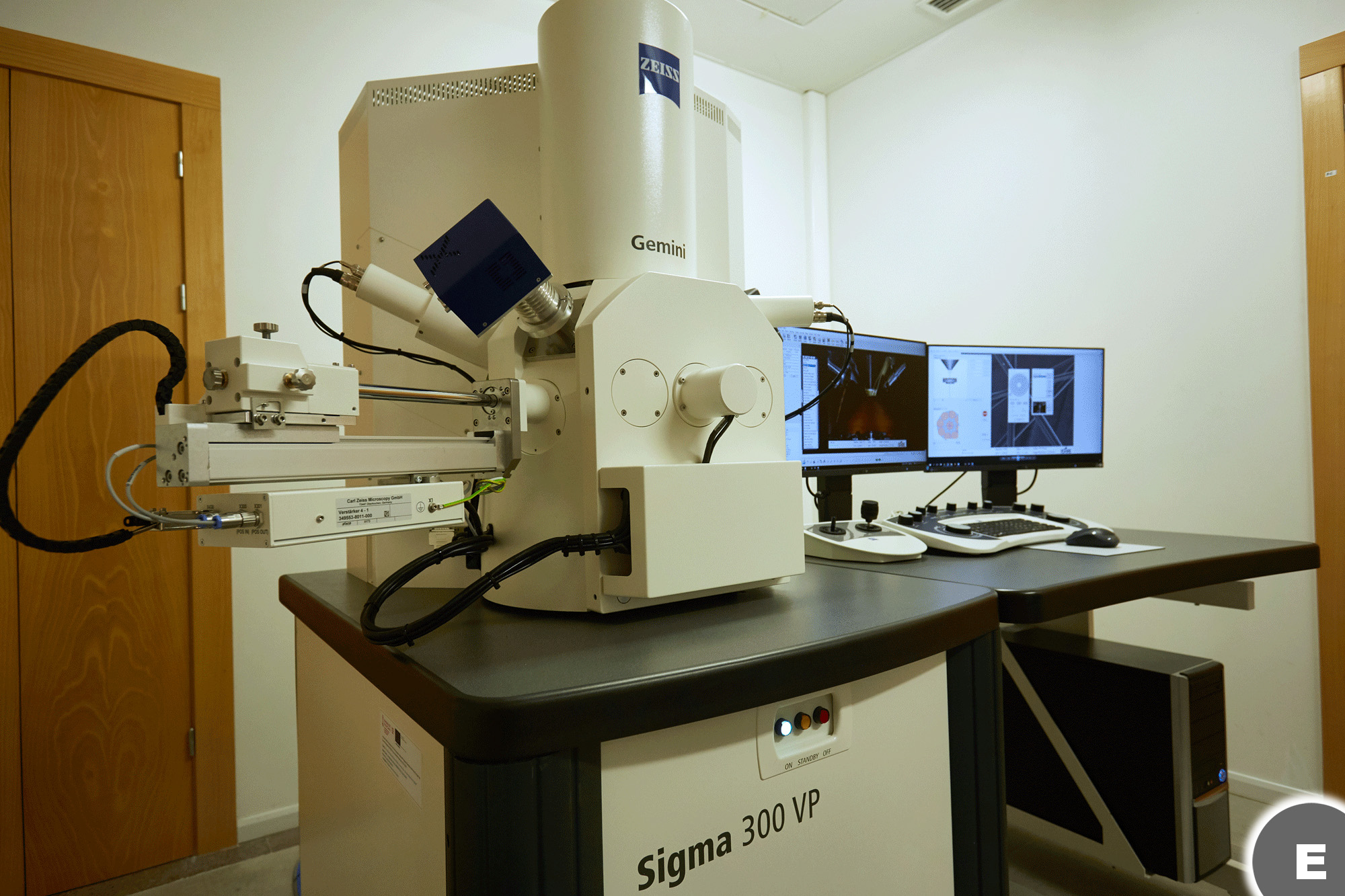 |
Microscopio electrónico de barrido de emisión de campo (FESEM) que permite la captura de imágenes de alta calidad combinada con el análisis elemental avanzado. |
|
|
|
||
|
Características destacadas:
|
||
Contacto
|
Campus de ELCHE Edificio TORREPINET |
Ver ubicación |
|
Tarifas vigentes (2026)
|
Referencia |
Descripción |
Unidad |
Precio unitario UMH (€) |
Precio unitario Entidades Públicas (€) |
Precio unitario Entidades Privadas (€) |
|
T-ELTP-04 |
Uso del microscopio electrónico de barrido de emisión de campo Zeiss Sigma 300 VP |
hora |
25.85 |
29.91 |
31.72 |
|
T-ELTP-07 |
Procesado de datos microscopio electrónico FESEM (Zeiss Sigma 300 VP) |
hora |
17.11 |
19.8 |
20.98 |
Observaciones
|
Tanto este microscopio electrónico como el microscopio óptico de barrido confocal del IDiBE pueden compartir el mismo tipo de portamuestras y hacen uso del mismo software, por lo que la microscopía correlativa CLEM (Correlative Light-Electron Microscopy) se puede llevar a cabo de una forma sencilla superponiendo y fusionando la información obtenida por ambas técnicas en una misma área de la muestra |
Financiación
|
El equipo ha sido adquirido gracias a una Actuación cofinanciada por la Unión Europea a través del Programa Operativo del Fondo Europeo de Desarrollo Regional (FEDER) de la Comunitat Valenciana 2014-2020 (PROYECTO: IDIFEDER2020/022). |
 |